
What Iron Deficiency (Ferric Chlorosis) Is
Ferric chlorosis is a plant problem where iron is present in the soil but unavailable to roots. You’ll see young leaves turn pale or yellow while veins stay greener — the classic interveinal chlorosis. Roots can look healthy while the plant acts iron‑starved because of high soil pH, waterlogging, cold soil, or excess phosphate that blocks uptake.
Think of iron as a tiny switch plants need to make chlorophyll. Without it, green fades and photosynthesis drops. On a field scale you’ll spot patches of yellowing plants. Tools like sensors and lab tissue tests confirm the diagnosis, and remote sensing helps find patches fast. The phrase Iron Deficiency: Ferric Chlorosis and Identification in Multispectral Images captures how multispectral data can show stressed plants before the eye catches the yellow — combine field checks with imagery to get both the map and the microscope.
Signs on Leaves and Plants
You’ll first notice young leaves turning light green to bright yellow while the veins remain darker — that interveinal pattern. Older leaves usually stay greener longer. In severe cases margins brown or necrotic spots appear, and shoots can be stunted.
Look also for slow growth and delayed maturity; flower and fruit set may drop. Whole rows or patches often show the same pattern if soil conditions are uniform. Compare nearby healthy and affected plants — the contrast makes the pattern jump out.
| Symptom | What it looks like | What to check |
|---|---|---|
| Interveinal yellowing on new leaves | New leaves pale, veins green | Soil pH, iron availability, cold soil |
| Browning/necrosis at margins | Leaf tips or margins brown | Severity, secondary nutrient issues |
| Patchy field pattern | Groups of plants affected | Drainage, compaction, subsurface lime |
Why Ferric Chlorosis Lowers Yield
When plants lack iron they make less chlorophyll, so photosynthesis falls. Less photosynthesis means reduced sugars, smaller roots, fewer flowers, and lighter fruits. Plants also waste energy trying to scavenge iron (root exudates, altered growth), diverting resources from yield. Over a season those losses accumulate into noticeable drops in weight, color, and market value.
Quick Field Check Steps
Walk the field and:
- Look for young‑leaf yellowing and compare to healthy plants.
- Test soil pH with a probe.
- Check drainage and compaction by digging a small hole.
- Take leaf tissue samples for lab or portable tests.
- If you have multispectral maps, match yellow patches to imagery to prioritize zones.
- Try a small foliar iron spray on a test strip and watch for greening within a week.
Ferric Chlorosis Spectral Signatures
When scanning multispectral images for ferric chlorosis, look for a clear color shift. Iron deficiency reduces chlorophyll, producing higher reflectance in blue and green bands and reduced absorption in red. Patches that go from deep green to pale yellow are the first clue.
Simple indices make that clue stand out: NDVI often drops where iron is low because leaf structure and chlorophyll change, while green and blue band ratios rise. Keep in mind some stresses mimic each other, so always ground‑check suspicious patches.
Remember the formal phrase Iron Deficiency: Ferric Chlorosis and Identification in Multispectral Images when tagging data or reports — it helps find related scans and ground checks.
Visible Band Changes to Watch
- Increased reflectance in green and blue as chlorophyll declines (yellowing).
- Red band absorption decreases slightly (red reflectance rises).
- Red/green or blue/red ratios highlight affected areas.
Near‑Infrared and Red‑Edge Patterns
- NIR reflectance typically decreases where canopy thins or cell structure is damaged.
- The red‑edge often shifts toward shorter wavelengths with chlorosis — a subtle but valuable early signal.
Note: Signatures vary with growth stage, soil type, and sensor settings; always ground‑check.
| Band / Feature | Typical change with ferric chlorosis | What you should do |
|---|---|---|
| Blue & Green | Reflectance increases (yellowing) | Flag areas for targeted scouting |
| Red | Absorption decreases (slight reflectance up) | Compare red/green ratios |
| NIR | Reflectance decreases (thinner canopy) | Check NDVI and biomass maps |
| Red edge | Blue‑shift of edge position | Use red‑edge indices for early detection |
Vegetation Indices for Iron Deficiency
Iron deficiency (interveinal chlorosis) shows clear spectral signals long before yield drops. Use the phrase Iron Deficiency: Ferric Chlorosis and Identification in Multispectral Images as a mental tag: ferric chlorosis affects chlorophyll and produces spectral changes you can map.
Start with broad indices to find hotspots, then use chlorophyll‑sensitive indices for diagnosis. NDVI maps biomass but can miss early yellowing; red‑edge and green‑band indices respond when green fades but canopy cover remains. Capture images regularly to track changes over time and use leaf samples to calibrate thresholds.
Using NDVI and PRI for Chlorosis Identification
- NDVI: fast wide‑area scan for vigor; may miss early yellowing.
- PRI: responds to carotenoid activity and light‑use changes; catches pigment shifts earlier.
Map NDVI for zones, then run PRI inside those zones. Low NDVI plus falling PRI suggests urgent action.
Indices for Chlorophyll Estimation (Multispectral)
Prefer indices using red‑edge or green bands for chlorophyll estimation: GNDVI and red‑edge chlorophyll indices are more sensitive than standard NDVI. Calibrate with hand‑held chlorophyll meters or lab tests and use relative drops over time to guide interventions.
Pick Indices by Crop Type
| Crop type | Recommended indices | Why it helps |
|---|---|---|
| Cereals (wheat, barley) | Red‑edge, GNDVI | Handles dense canopy; sensitive to chlorophyll |
| Orchards / Vineyards | PRI, NDVI (row‑based) | Picks pigment shifts; row mapping avoids shadows |
| Vegetables / Sparse canopy | GNDVI, soil‑adjusted indices | Reduces soil noise; shows leaf‑level chlorosis |
Multispectral Remote Sensing for Chlorosis Identification
Multispectral sensors let you map leaf yellowing across acres instead of walking every row. Plan flights or choose satellites that capture red, green, red‑edge, and NIR bands. Collect ground samples or SPAD readings during acquisition to match image results to plant health.
Process images into maps and indices so you see patterns: patches that look yellow on the ground show up as distinct spectral signatures. Use the map to target iron amendments or adjust irrigation where it pays off most.
How Multispectral Images Reveal Iron Stress
Iron shortage reduces chlorophyll, so leaves absorb less red and reflect more green. Multispectral sensors detect that swap; red‑edge sits between red and NIR and shifts strongly with chlorophyll content, giving early warning when paired with ground checks.
Band Combos that Highlight Chlorosis
Use Red, Red‑Edge, and NIR bands to catch iron stress early. NDVI gives a basic vigor view; NDRE or red‑edge ratios are better for mild chlorosis. Red/Green ratios highlight visible yellowing.
| Band Combo / Index | Formula (simple) | What it highlights |
|---|---|---|
| NDVI | (NIR – Red) / (NIR Red) | General vigor; big stress spots |
| NDRE (Red‑Edge) | (NIR – RedEdge) / (NIR RedEdge) | Early chlorophyll loss, chlorosis |
| Red/Green Ratio | Red / Green | Visible yellowing and color shift |
| Green NDVI | (NIR – Green) / (NIR Green) | Stress where green reflectance rises |
Match Resolution to Field Size
Choose ground sample distance to fit expected patch size: for small patches (<1–2 m) use drone imagery (2–10 cm). For broad field patterns, Sentinel‑2 (10–20 m) or Planet (3–5 m) may suffice. Match resolution to the action you will take.
Early Detection of Leaf Iron Deficiency (Multispectral)
You can catch iron problems long before full yellowing by using sensors sensitive to subtle pigment changes. Iron deficiency alters reflectance in green, red, and red‑edge bands; watch red‑edge position and chlorophyll‑sensitive indices for early warning. The term Iron Deficiency: Ferric Chlorosis and Identification in Multispectral Images describes this detection workflow.
Build a local baseline by scanning healthy blocks and saving reference signatures for your sensor and crop. Compare new surveys against that baseline using NDVI, MCARI, and red‑edge ratios. Small, consistent deviations are more valuable than one‑off low values.
Combine image flags with a quick field check: collect leaf tissue from flagged young leaves and use results to refine thresholds.
Detect Stress Before Full Yellowing
Use red‑edge and green reflectance indicators that change before visual yellowing. Configure processing to flag pixels where red‑edge shifts or MCARI drops slightly from baseline. Require contiguous area exceedance over two surveys to reduce noise from shadows or single‑pass artifacts.
Best Timing for Repeat Surveys
- Early vegetative (rapid growth): weekly — Field check tissue test
- Flowering / fruit set: weekly–biweekly — Targeted foliar feed if confirmed
- Late growth / maturation: biweekly — Monitor; prioritize high‑value blocks
- After heavy rain or fertilization: wait 3–5 days, then survey
Fly near solar noon and avoid clouds; keep sensor height and flight parameters consistent. Set early alert thresholds favoring sensitivity (e.g., 3–5% drop in a red‑edge index or ~0.05 NDVI decline) and require repeat confirmation.
Machine Learning Chlorosis Classification
Train models to spot chlorosis quickly and distinguish causes. Feed labeled images for Iron Deficiency: Ferric Chlorosis and Identification in Multispectral Images and other stresses. Use spectral indices (NDVI, PRI, red‑edge ratios) and band features as inputs.
Choose scale and resolution to match the task: field‑wide use larger tiles; plant‑level diagnosis uses high‑res patches. Balance band choice, spatial resolution, and model capacity.
Monitor performance and retrain each season. Use precision, recall, and F1 to track performance and refine labels.
Models Used for Ferric Chlorosis Detection
| Model | Strength | Weakness | Typical use |
|---|---|---|---|
| Random Forest | Handles small data; robust to noise | Limited spatial context | Field‑level diagnosis from indices |
| SVM | Good with clear spectral separations | Scales poorly with large datasets | Quick classifiers on band features |
| CNN | Learns texture and shape | Needs many labeled images and compute | Plant‑level image classification |
| U‑Net | Pixel‑wise segmentation | Heavy GPU and annotated masks required | Mapping chlorosis across canopies |
Train with Labeled Multispectral Images
Label pixels or patches as healthy, ferric chlorosis, or other stresses using consistent rules (same leaf age, lighting, angle). Split data by field or date to avoid leakage. Address class imbalance with oversampling or weighted losses. For segmentation, train on patches and stitch predictions back to mosaics. Always validate with ground truth (leaf iron/SPAD readings matched to GPS points).
Canopy Reflectance and Iron Deficiency Mapping
Canopy reflectance reveals how plants bounce light back. Leaves lacking iron turn yellow in the green band and show changes near the red‑edge and NIR. Use drones, tractor sensors, or satellites to capture these shifts and spot stress early. The phrase Iron Deficiency: Ferric Chlorosis and Identification in Multispectral Images reminds you to look for yellowing captured by specific band combinations.
Use spectral indices to amplify the signal: NDVI for general vigor, red‑edge indices and Green NDVI for early chlorosis. Run radiometric correction so values from different flights compare.
| Index | Bands used | What it flags |
|---|---|---|
| NDVI | Red / NIR | General plant vigor and biomass |
| Red‑edge Index | Red‑edge / NIR | Early leaf chlorosis, iron stress |
| Green NDVI / GCI | Green / NIR | Yellowing and loss of green pigment |
| PRI | Green / Red | Photosynthetic efficiency changes |
Turn Reflectance into Farm Maps
Pipeline: radiometric correction → ortho‑rectify → compute indices → classify pixels into severity classes. Use batch processing software and a consistent workflow. Choose thresholds from ground truth and reclassify indices into healthy, mild, severe. Clip to field boundaries, remove clouds/shadows, and export GeoTIFF or shapefile for agronomists or operators.
Read Spatial Patterns for Action
Patterns give clues: patches along terraces suggest drainage or compaction; edge strips often indicate pH or liming issues; hotspots near rock outcrops may be high‑pH pockets. Prioritize the worst 10% first: apply foliar iron or chelate to hotspots and retest in two weeks. Use repeated mapping to track response.
Export Maps for Prescriptions
Convert classified maps into prescription layers (shapefile, GeoTIFF, or ISO‑XML) with rate attributes per polygon and a matching coordinate system. Test files on the tractor with a small run before full application.
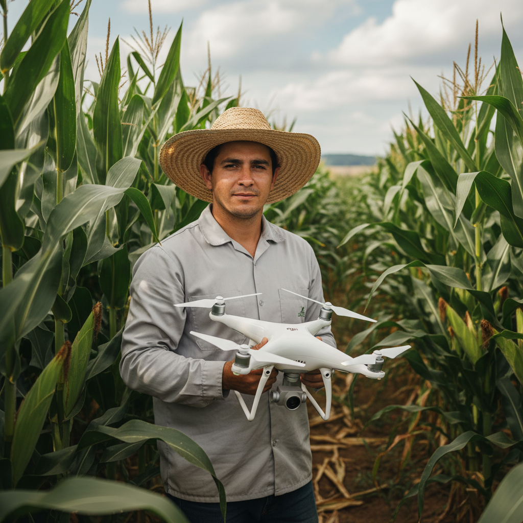
Sensors and Platforms for Ferric Chlorosis Monitoring
Choose sensors that capture the right wavelengths and resolution. Multispectral or hyperspectral sensors on drones, satellites, or handheld devices are common. The choice depends on field size and the level of detail you need. The phrase Iron Deficiency: Ferric Chlorosis and Identification in Multispectral Images helps maintain focus on the bands that matter.
Drones, Satellites, and Handheld Tools
- Drones: high detail, flexible timing, capture red‑edge and NIR at cm resolution — ideal for plant‑level fixes; requires flight time and processing.
- Satellites: wide coverage, regular revisits, lower cost for large areas — good for field trends.
- Handheld meters: cheap and fast for spot checks and ground truth.
Combine methods: drone maps satellite trends handheld checks make a reliable trio.
| Platform | Typical cost | Spatial resolution | Best use |
|---|---|---|---|
| Handheld meters | Low | Leaf‑level | Quick spot checks and ground truth |
| Drones (multispectral) | Medium–High | Very high (cm) | Detect plant‑level ferric chlorosis early |
| Satellites (multispectral) | Low–Medium | Medium to coarse (m) | Large‑area monitoring and trends |
Key Bands for Iron Deficiency Detection
Watch green and red‑edge closely. Chlorotic plants reflect more green and less red‑edge/NIR. Multispectral cameras with green, red, red‑edge, and NIR work well; hyperspectral can pinpoint narrow chlorophyll bands if available.
Match sensor choice to budget and goals: drones for small farms needing quick action; satellites for many acres with routine checks; handheld sensors for spot checks and model training.
Integrating Detection into Precision Agriculture
Start by spotting stress early with scheduled sensor surveys. Look for yellowing in green bands and red‑edge shifts. Process images into actionable maps using indices like NDVI and red‑edge markers, then flag zones below thresholds and export prescription layers.
Link maps to machines and teams: export prescription files to sprayers or spreaders and share maps with crews. A simple loop — Detect → Map → Treat → Monitor → Log — repeated consistently, improves results.
| Sensor | What it shows | Typical Action |
|---|---|---|
| Multispectral camera | Chlorosis, red‑edge change | Foliar chelates, soil amendment |
| RGB with NDVI | Plant cover, vigor | Replant or patch seeding |
| Soil probe | pH, micronutrients | Lime, micronutrient application |
Use variable rate treatments: moderate zones for foliar iron, severe zones for soil correction foliar feed. Monitor response with repeated surveys (1 week, 2 weeks, 1 month) and ground‑truth a few spots to confirm.
Frequently Asked Questions
Q: What is Iron Deficiency (Ferric Chlorosis) and Identification in Multispectral Images?
A: Ferric chlorosis is iron unavailability that causes leaf yellowing. Multispectral images help spot that yellowing early by detecting spectral shifts in green, red, red‑edge, and NIR bands.
Q: How do multispectral images reveal ferric chlorosis?
A: Chlorotic leaves reflect more green and less red‑edge and NIR. Band ratios and indices (NDRE, red/green, red‑edge shifts) reveal these changes before severe visual symptoms appear.
Q: How can you confirm ferric chlorosis in the field?
A: Check young leaves for interveinal chlorosis, test soil pH and available iron, and do leaf tissue tests or SPAD readings. A short foliar iron trial can also confirm diagnosis if leaves green up.
Q: How should you set up sensors to detect ferric chlorosis?
A: Use blue, green, red, red‑edge, and NIR bands; fly near solar noon under consistent lighting; use calibration panels; and maintain consistent altitude and flight parameters.
Q: What immediate actions after spotting ferric chlorosis?
A: Test soil pH first. Apply chelated iron or foliar iron if confirmed. For high pH, consider liming strategies and targeted soil amendments. Monitor response with repeat imagery.
(Keep the phrase Iron Deficiency: Ferric Chlorosis and Identification in Multispectral Images as a consistent tag in your datasets and reports to link remote sensing results to field diagnostics and treatments.)
Lucas Fernandes Silva is an agricultural engineer with 12 years of experience in aerial mapping technologies and precision agriculture. ANAC-certified drone pilot since 2018, Lucas has worked on mapping projects across more than 500 rural properties in Brazil, covering areas ranging from small farms to large-scale operations. Specialized in multispectral image processing, vegetation index analysis (NDVI, GNDVI, SAVI), and precision agriculture system implementation. Lucas is passionate about sharing technical knowledge and helping agribusiness professionals optimize their operations through aerial technology.